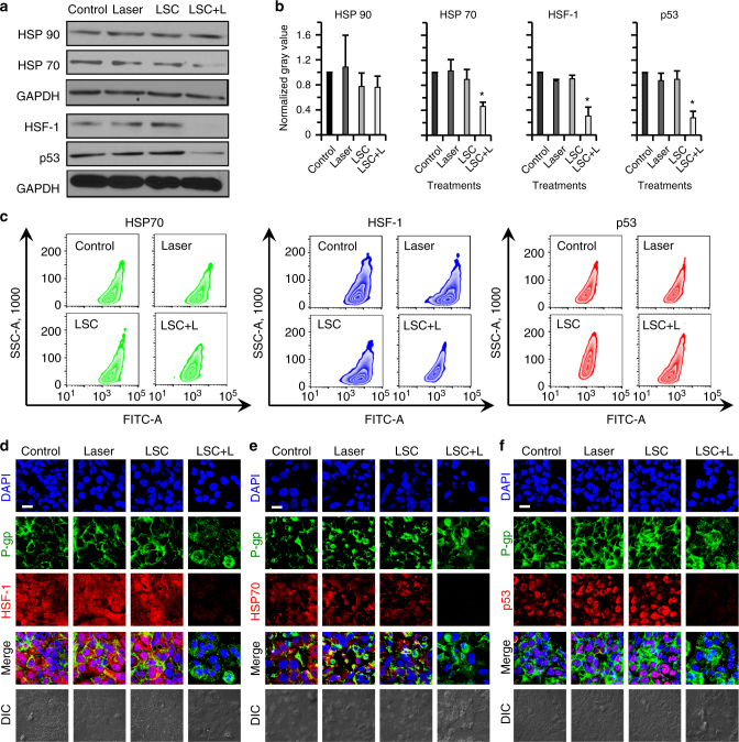
Fig. 8.
Possible mechanisms of the LSC + L treatment in sensitizing the drug-resistant cells to cancer therapy. a Qualitative western blot data of heat shock protein 90 and 70 (HSP90 and HSP70), heat shock factor-1 (HSF-1), and mutant p53 proteins, showing decreased expression of HSP70, HSF-1, and mutant p53 after the treatment of LSC nanoparticles and NIR laser irradiation (LSC + L). b Quantitative data of the western blot results, showing the decreased expression of HSP70, HSF-1, and mutant p53 in the NCI/RES-ADR cells with the LSC + L treatment is statistically significant. Error bars represent s.d. (n = 3). *p < 0.05 (Kruskal–Wallis H-test). c Flow cytometry analysis showing decreased expression of HSP70, HSF-1, and mutant p53 in NCI/RES-ADR cells treated with LSC nanoparticles and NIR laser irradiation. d Confocal images of HSF-1 showing the HSF-1 distributes in both nuclei and cytoplasm. e Confocal images of HSF70 showing the HSF70 mainly distributes in cytoplasm. f Confocal images of mutant p53 show the mutant p53 mainly distributes in nuclei. The distribution of P-gp is similar to that shown in Fig. 4a. No treatment (laser or nanoparticle) was conducted on the cells in the control group. The cells were permeabilized for the flow cytometry and confocal analyses. The NIR laser irradiation was at 1 W cm−2 for 1 min. The concentration of LSC nanoparticles used for the LSC or LSC + L groups was 0.2 mg ml−1. Scale bars: 20 μm

