Fig. 1.
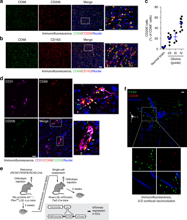
Alternatively activated macrophages are localized proximately to GBM-associated ECs. a–d Tissue sections from human normal brain and surgical specimens of human glioma tumors were probed with different antibodies. a GBM tumor sections were stained with anti-CD68 and anti-CD206 antibodies. Representative images are shown (n = 5 GBM patient tumors). Bar represents 100 μm. Zoom-in factor: 4. b GBM tumor sections were stained with anti-CD68 and anti-CD163 antibodies. Representative images are shown (n = 5 patient GBM tumors). Bar represents 100 μm. Zoom-in factor: 4. c Normal brain and GBM tumor sections were stained with anti-CD68 and anti-CD206 antibodies. Quantified data are shown (total n = 4 normal brains and 21 glioma tumors, mean ± SEM). d GBM tumor sections were stained with anti-CD31, anti-CD206, and anti-CD68 antibodies. Representative images are shown (n = 5 patient GBM tumors). Arrows indicate CD68+CD206+ cells. Bar represents 100 μm. Zoom-in factor: 4. e, f GBM was induced by RCAS-mediated gene transfer in Ntv-a;Ink4a-Arf−/−;Ptenfl/fl;LSL-Luc mice, followed by orthotopic tumor transplantation into Rosa-LSL-tdTomato;Tie2-Cre mice. e Experimental procedure. f Thick sections were stained with anti-F4/80 and anti-CD206 antibodies, and subjected to confocal scanning imaging. 3-D images were generated and shown. Bar represents 200 μm. Zoom-in factor: 1.6

