Fig. 7.
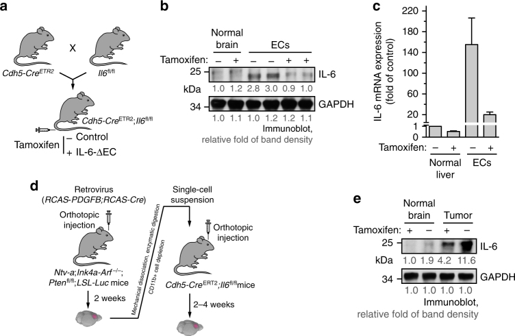
ECs are a major source for IL-6 expression in GBM. a–c Cdh5-CreERT2;Il6fl/fl mice were generated by crossing Cdh5-CreERT2 mice with Il6fl/fl mice. Mice (2 weeks old) were injected with tamoxifen for consecutive 5 days to induce EC-specific IL-6 knockout. a Schematic approach. b, c ECs were isolated from mouse aortas. b Brain tissue and ECs were subjected to immunoblot analysis. Band density was quantified. c mRNA was extracted and subjected to quantitative RT-PCR analysis. Results were normalized with GAPDH level and expressed as folds of control (n = 3, mean ± SEM). d, e The primary GBM in Ntv-a;Ink4a-Arf−/−;Pten−/−;LSL-Luc donor mice was induced by RCAS-mediated somatic gene transfer. Recipient mice were Cdh5-CreERT2;Il6fl/fl mice. d Schematic approach. e Normal brain and tumor tissues were homogenized. Tissue lysates were immunoblotted. Band density was quantified

