Fig. 8.
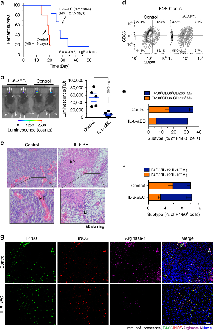
Endothelial IL-6 is critical for macrophage alternative activation and GBM growth and progression. The genetically engineered GBM model was induced in Ntv-a;Ink4a-Arf−/−;Pten−/−;LSL-Luc donor mice, followed by orthotopic tumor implantation into Cdh5-CreERT2;Il6fl/fl mice that were treated with (IL-6-ΔEC) or without (Control) tamoxifen. a Animal survival was monitored for 50 days post-injection (n = 5–6 mice, one representative result from three independent experiments). P values were determined by log-rank (Mantel–Cox) tests. MS, median survival. b Tumor growth was analyzed by bioluminescence. Left, representative images. Right, quantitative analysis of integrated luminescence in tumors at day 12 (mean ± SEM, n = 5–6, one representative result from three independent experiments). P value was determined by Student’s t test. c Tumor sections were stained with hematoxylin and eosin (H&E). Representative images are shown (n = 10 mice). P pseudopalisades, MP microvascular proliferation, EN extensive necrosis, LI leukocyte infiltration. Bar represents 100 μm. Zoom-in factor: 3. e, f Tumors were excised. Single-cell suspensions were prepared and subjected to flow cytometry analysis. d, e Single-cell suspensions were probed with anti-F4/80, anti-CD86, and anti-CD206 antibodies. CD206 and CD86 expression were analyzed in sorted F4/80+ cells. d Representative sorting. e Quantified results (mean ± SEM, n = 10–14 mice). f Single-cell suspensions were probed with anti-F4/80, anti-IL-10, and anti-IL-12 antibodies. IL-10 and IL-12 expression was analyzed in sorted F4/80+ cells. Show are quantified results (mean ± SEM, n = 8–13 mice). g Tumor sections were stained and analyzed by immunofluorescence. Tumor sections were probed with anti-iNOS, anti-arginase-1, anti-F4/80 antibodies (n = 10 mice). Bar represents 100 μm

