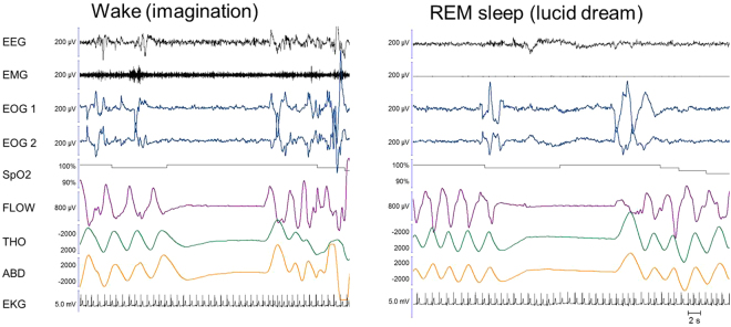
Figure 2.
Example of an apnoea executed during an imagined diving task during wakefulness (left panel) and in a lucid dream during coded REM sleep (right panel). In the right panel, the absence of muscle tone in the EMG and the theta frequency on the EEG confirmed that the patient was in REM sleep during the lucid dream. Between the two ocular left-right codes, we observe an 11-second-long central apnoea, characterized by a cessation of nasal flow along with the absence of thoracic and abdominal movements and a mild heart rate decrease. After this episode, the patient reported a dream in which he was threatened by a gun and held his breath in fear. Note that in the imagined condition (left panel), the last breath before the apnoea appears slightly different from the previous ones regarding its time dynamics, but there is no evidence of augmented amplitude. In the lucid dream condition, there is no evidence of pre-apnoeic preparation. EEG: electro-encephalogram; EMG: electromyogram; EOG1 and EOG2: electro-oculogram for left and right movements, respectively; SpO2; transcutaneous oxygen saturation; FLOW: nasal flow; THO: thoracic movements; ABD: abdominal movements; EKG: electro-cardiogram

