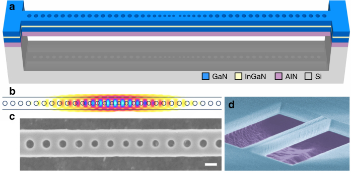
Fig. 1.
III-nitride nanobeam cavity membrane. a Schematic drawing of a freestanding nanobeam structure featuring a single InGaN/GaN QW. It consists of two photonic crystal mirrors tapered down to the cavity (not drawn to scale for clarity). b Field intensity profile |Ey|2 of the fundamental cavity mode as obtained via 3D-FDTD simulations. c, d Top and side-view scanning electron microscope images of typical nanobeam structures, where the III-nitride layer, the airgap, and the silicon underneath (false-color in d) are noticeable. The scale bar in c is equal to 100 nm in length

