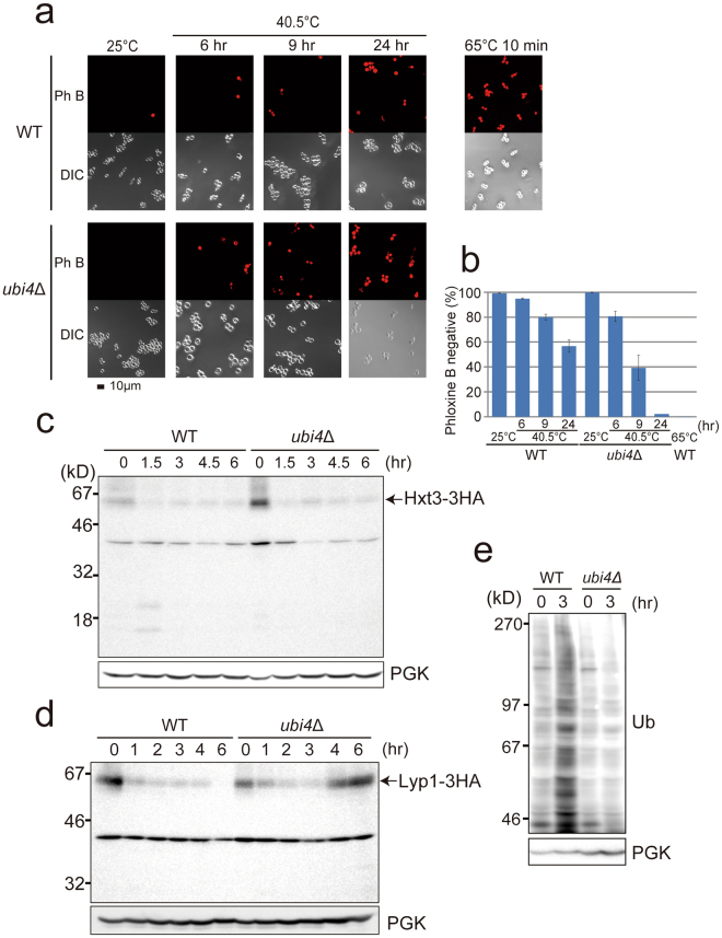
Figure 1.
Physiological properties of cells under chronic heat stress in wild-type and ubi4Δ cells. (a) Phloxine B staining. Wild-type W303 and ubi4Δ cells grown at 25 °C were transferred to 40.5 °C, or wild-type cells incubated at 65 °C for 10 min and Phloxine B-stained cells were observed by fluorescence microscopy. Dead cells were stained by Phloxine B. (b) Quantification of Phloxine B-unstained cells in (a). Date are presented as the mean value of three independent experiments, except for the value of ubi4Δ cells at 40.5 °C for 24 h, which is an average of two experiments. Standard errors (SE) are shown. (c–e) Lyp1-3HA, Hxt3-3HA and ubiquitin accumulations in wild-type and ubi4Δ cells at the indicated times after exposure to 40.5 °C. Anti-HA antibody for Lyp1-3HA and Hxt3-3HA. Anti-phosphoglycerate kinase (PGK) antibody as a control for protein loading. Molecular weight markers are indicated. The levels of ubiquitinated proteins were lower in ubi4Δ cells than in wild-type cells after 3 h at 40.5 °C.

