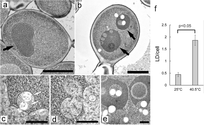
Figure 4.
Electron microscopy of wild-type cells, and lipid droplet formation after chronic heat stress. (a) Wild-type cells at 25 °C. (b) Wild-type cells grown at 40.5 °C for 6 h. (c–e) Various types of vacuolar invaginations in cells grown at 40.5 °C for 6 h. Scale bar; 2 μm for (a) and (b), and 0.5 μm for (c–e). Black and white arrows indicate vacuoles and MVBs, respectively. (f) Number of lipid droplets per a cell at 25 °C and 40.5 °C for 6 h. Average were calculated from EM images of fifty-two cells that were randomly taken. Error bar indicates SE, and significant difference was calculate using Student’s-t test.

