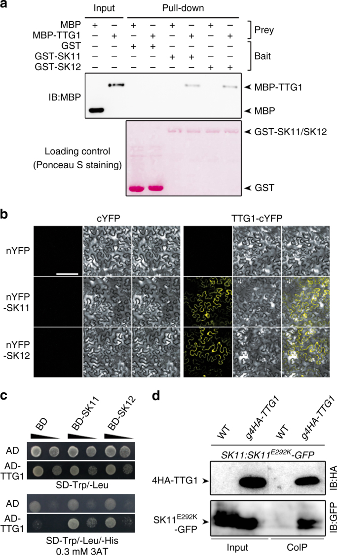
Fig. 1.

SK11 and SK12 interact with TTG1. a GST pull-down assay of the interaction of TTG1 with SK11 and SK12. GST, GST-SK11, and GST-SK12 were used as baits, and their loading amounts stained with Ponceau S are shown in the bottom panel. The input of prey proteins, MBP and MBP-TTG1, and their corresponding pull-downed signals are shown in the upper panel. b BiFC analysis of the interaction between TTG1 and SK11/SK12 in tobacco epidermal cells. Scale bar, 200 μm. c Yeast two-hybrid assay of the interaction between TTG1 and SK11/SK12. Ten-fold serial dilutions of transformed yeast cells were grown on SD-Trp/-Leu medium (upper panel) and SD-Trp/-Leu/-His medium supplemented with 0.3 mM 3-amino-1, 2, 4-triazole (3AT) (lower panel). d In vivo interaction between 4HA-TTG1 and SK11E292K-GFP in Arabidopsis siliques shown by co-immunoprecipitation (CoIP). Total protein extracts from siliques of SK11:SK11E292K-GFP in wild-type (WT) or g4HA-TTG1 background 4 days after pollination (DAP) were immunoprecipitated by anti-HA antibody. The input and co-immunoprecipitated proteins were subjected to immunoblot analysis with anti-HA (upper panel) or anti-GFP antibody (lower panel). Uncropped original scans of immunoblots are shown in Supplementary Fig. 17
