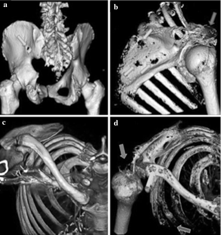
Fig. 2.
3D reconstructions of computerised tomography (CT) images using standard diagnostic settings demonstrating two patients with widespread myeloma-induced bone disease, leading to potential serious consequences. a Lytic lesion penetrating through the ischium (green arrow). b Multiple lytic lesions throughout the scapula (green arrows) with the acromion completely destroyed by myeloma bone disease (red arrow). c Example of normal bone from the shoulder, clavicle and ribs. d Contrast image of the patient riddled with lytic lesions due to myeloma bone disease. The acromion process is destroyed (red arrow), multiple lytic lesions are present throughout the clavicle (green arrow) and the anterior ribs have been destroyed (purple arrow) (Color figure online)

