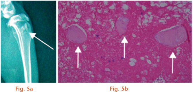
a) Typical radiological evidence of chronic osteomyelitis: reduced bone density; dead bone; subperiosteal abscesses; and new periosteal bone formation (white arrow: sequestrum formation). b) The haematoxylin and eosin staining showed a large number of clearly visible inflammatory cells, interstitial haemorrhage and bone sequestrum formation (white arrow).
