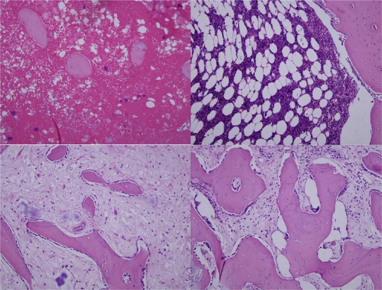
Fig. 7.
Histological observation for the repair of osteomyelitis defects in different groups at eight weeks after implantation. (Hematoxylin-eosin stain × 100) Control group (top left): many inflammatory cells were observed, accompanied by sequestrum formation. G-TCP0 group (top right): most of the composite scaffold was clearly visible and no new bone had formed. G-TCP1 group (bottom left): only small amount of material was visible and some islands of new trabecular bone had formed. G-TCP3 group (bottom right): the composite scaffold was completely degraded and a large amount of new bone had formed.

