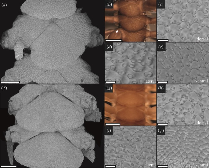
Figure 2.
Calcite elements on the arm plates in Ophiocoma echinata and O. pumila visualized by synchrotron X-ray tomography (a, c–f, h–j) and photography (b,g). Ophiocoma echinata (a–e) is covered with very regular, hemispherical EPTs on the dorsal arm plates (a–c), ventral arm plates (d), and the dorsal and ventral regions of the lateral (a,e) arm plates. The EPTs are surrounded by pigmented chromatophores giving a dark colour (b). Ophiocoma pumila (f–j) lacks chromatophores and appears much paler (g). The skeletal elements are less regular than the EPTs observed in O. wendtii (figure 1) and O. echinata (a–c), but EPT-like hemispheres are present across the dorsal arm plates (f,h), margins of the lateral arm plates (i) and ventral arm plates (j). See the electronic supplementary material (S2–S3) for reconstructed models. Scale bars: (a,f) 250 µm; (b,g) 500 µm; (c–e, h–j) 25 µm. (Online version in colour.)

