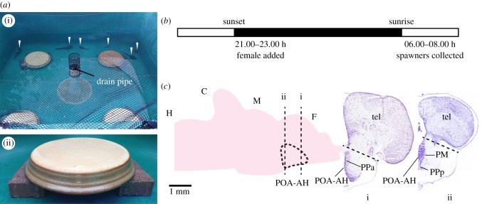
Figure 1.
Experiment design (a) Overhead view of behaviour tank with four nests (i) with non-nesting males visible (arrows), and close up of individual nest (ii). (b) Timeline for spawning trials. (c) Left: sagittal view of midshipman brain. Preoptic area-anterior hypothalamus (POA-AH) highlighted in blue. Right: coronal sections through midshipman forebrain at the level of the dashed lines labelled i and ii through POA-AH in left panel. Dashed lines indicate level of cuts separating POA-AH and telencephalon during dissection. Stained with cresyl violet. H, hindbrain; C, cerebellum; M, midbrain; F, forebrain; PM, magnocellular preoptic area; PPa, anterior parvocellular preoptic area; PPp, posterior parvocellular preoptic area; Tel, telencephalon.

