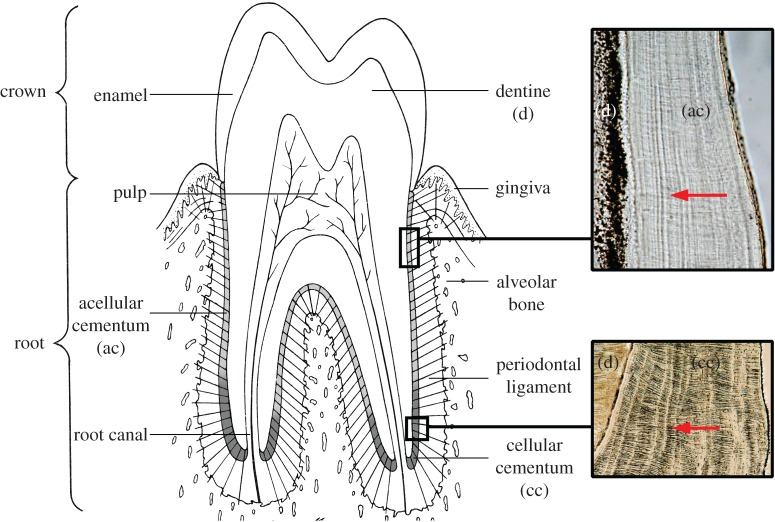
Figure 1.
The tissue components of a tooth and periodontium showing the location of acellular (ac) and cellular cementum (cc) on the surface of the root dentine (d). Red arrows within boxes show the direction of extrinsic fibres, more prominent in (cc), also known as Sharpey's fibres. Bright incremental markings in both (ac) and (cc) run near-vertically. (Online version in colour.)

