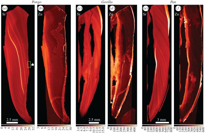
Figure 2.
Overview SXRF scans (25 µm) of Sr and Zn distribution in the incisor tooth roots of Pongo (UCL-JS3-CA-28-UI1), Gorilla (UCL-CA1G-1474-UI1) and Pan (UCL-CA-14E-LI1). Secondary dentine (on the pulpal aspect of each root) is to the left of each pair of images and cementum (on the root surface) to the right of each pair. Sr concentration (a,c,e) is extremely variable, but generally follows the incremental growth pattern of root dentine. Zinc concentration (b,d,f) is highest in the secondary dentine surrounding the pulp cavity and in the cementum layers. The region highlighted by the green box in (a) in the Sr map (⋆) identifies regular compensatory acellular cementum scanned at 5 µm shown in figure 3. (Online version in colour.)

