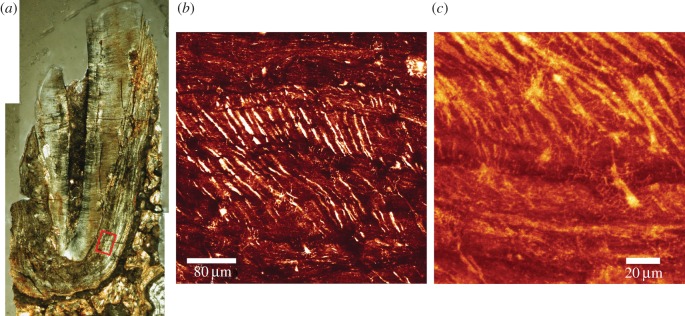
Figure 6.
TLM (a) of canine root of KNM-ER 1817. Laser confocal image (b) of cementum layers, taken within the region of the red box. The right border of the red box is the lower border of the middle and right fields of view. Air-filled Sharpey's fibre spaces change orientation as they cross cementum layers. At higher magnification cementocyte lacunae and their canaliculi can also be imaged in this fossil (c). (Online version in colour.)

