Figure 4.
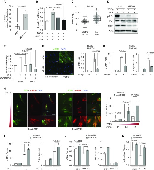
PDK1 promotes myofibroblast differentiation. (A) Lactate concentration of lung tissue from PBS- and bleomycin-treated mice (n = 4 per group). Two-tailed t test. (B) Extracellular lactate measurement in siScr or siHIF-1α IPFs treated with TGF-β or TGF-β + dichloroacetate (DCA; n = 6 from three biologically independent experiments). IPFs were treated with 2 ng/ml TGF-β and 10 mM DCA for 72 hours. (C) Microarray gene profile analysis of PDK1 expression between control subjects (n = 137) and patients with interstitial lung disease (ILD; n = 255), derived from the Lung Genomics Research Consortium database. Mann-Whitney U test. (D) Western blot analysis of p-pyruvate dehydrogenase (PDH), total PDH, and PDK1 expression in siScr and siPDK1 HLFs treated with 5 ng/ml TGF-β and 10 mM DCA for 24 hours. These results were observed from three independent experiments. (E) Extracellular lactate measurement in siScr or siPDK1 HLFs treated with TGF-β or TGF-β + DCA (n = 6 from three biologically independent experiments). IPFs were treated with 2 ng/ml TGF-β and 10 mM DCA for 48 hours. (F) Representative immunocytochemistry and quantification of α-SMA in siScr and siPDK1 HLFs treated with 2 ng/ml TGF-β for 72 hours (n = 6 from three biologically independent experiments, 5–10 images were captured per group and normalized to nuclei for quantification). Two-tailed t test. (G) qRT-PCR analysis of α-SMA and calponin1 mRNA expression in siScr and siPDK1 HLFs treated with 2 ng/ml TGF-β for 72 hours (n = 4 from two biologically independent experiments). Two-tailed t test. (H) Representative immunocytochemistry and quantification of α-SMA in lentivirus (lenti)-GFP and lenti-PDK1 HLFs treated with the indicated TGF-β concentrations for 72 hours (n = 6 from three biologically independent experiments, 5–10 images were captured per group and normalized to nuclei for quantification). (I) qRT-PCR analysis of α-SMA and calponin1 mRNA expression in lenti-GFP and lenti-PDK1 HLFs treated with 2 ng/ml TGF-β for 72 hours (n = 4 from two biologically independent experiments). Two-tailed t test. (J) qRT-PCR analysis of α-SMA and HIF-1α expression in siScr/lenti-GFP or lenti-PDK1, and siHIF-1α/lenti-GFP or lenti-PDK1 HLFs treated with 2 ng/ml TGF-β for 72 hours (n = 8 from four biologically independent experiments). (K) Extracellular lactate measurement in siScr/lenti-GFP or lenti-PDK1, and siHIF-1α/lenti-GFP or lenti-PDK1 HLFs treated with 2 ng/ml TGF-β for 72 hours (n = 8 from four biologically independent experiments). Scale bars: 100 μm. Error bars represent the mean (±SEM). One-way ANOVA with multiple comparison, post hoc, unless otherwise noted.

