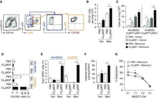
Figure 2.
Infiltration of myeloid-derived suppressor cells (MDSCs) expressing CXCR2 into the lung in bleomycin treated mice with macrophage depletion. (A) Representative flow plot, where CD11b+Ly6ChiLy6G- cells (highlighted in blue) represent monocytic MDSCs (Mo-MDSCs), and CD11b+Ly6CloLy6G+ cells (highlighted in orange) represent granulocytic MDSCs (G-MDSCs). Gated on live singlet CD45+ cells. (B) The absolute cell numbers of CD11b+ cells, determined based on lung cellularity. (C) The absolute cell numbers of Mo-MDSC and G-MDSC of CD11b+ cells based on lung cellularity. (D) The percentage CXCR2+ cells of Mo-MDSC or G-MDSC cell population. (E) Percent arginase (Arg) 1+ inducible nitric oxide synthase (iNOS)+ cells in Mo-MDSCs and G-MDSCs. (F) Whole-lung Arg activity of liposome-treated wild-type mice (n = 12 per group over course of three independent experiments). (G) CD11b+Gr-1+ cells were purified from the spleens of bleomycin- and liposome-treated mice and placed in a suppression assay with purified carboxyfluorescein succinimidyl ester–stained C57BL/6 T cells at the designated ratios. Anti-CD3/CD28 beads were used to induce proliferation. Cells were harvested on Day 5 and analyzed by flow cytometry for dilution (n = 8 per mouse group experiment). Results are plotted as the mean (±SEM). *P < 0.05. Ly6 = lymphocyte antigen 6 complex.

