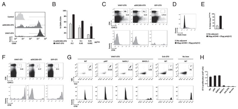
FIGURE 4.
Targeting of MHCII products via VHH7 fusions elicits strong CD4 T cell proliferation. (A–C) At equimolar concentrations, VHH7-OTII (0.2 μg) is more efficient at activating specific CD4+ T cells than is anti-DEC205–OTII (1 μg), in vitro and in vivo. (A and B) DCs were incubated with OTII-specific CD4 T cells from OT-II–transgenic mice, and T cell activation was monitored by CD69 cell surface expression. (A) Graphs represent CD69 staining of OT-II T cells after incubation of DCs with equimolar concentrations of VHH7-OTII or anti-DEC205–OTII. (B) Quantification of CD69 surface expression in OT-II T cells after incubation of DCs with different concentrations of VHH7-OTII or anti-DEC205–OTII. (C) CFSE-labeled CD4+ T cells from CD45.2 OT-II–transgenic mice were transferred i.v. into CD45.1 C57BL/6 mice (n = 3 per group). One day later, recipient mice were left untreated or were immunized with equimolar amounts of VHH7-OTII, anti-DEC205–OTII, or GFP-OTII in solution with 25 μg of anti-CD40 and 50 μg of poly(I:C). CD4 T cell expansion was measured by flow cytometry 7 d later. Staining of splenocytes with fluorescently labeled anti-CD4 and anti-CD45.2 shows stronger proliferation of anti-OTII CD4 T cells in VHH7-OTII–immunized mice. Graphs of donor OT-II CD45.2 cells show loss of CFSE labeling in proliferating cells. (D and E) In vivo targeting of VHH7-OTII with anti-CD40 and poly(I:C) induces stronger proliferation of OT-II CD4 T cells than does VHH7-OTII alone. (D) CellTrace Violet–labeled CD4+ T cells from CD45.2 OT-II–transgenic mice were transferred i.v. into CD45.1 C57BL/6 mice (n = 3 per group). Immunization and readout are as in (C). (E) Quantification of CellTrace Violetlow/neg OT-II T cells in (D). (F) VHH7-OTI fails to activate CD8+ T cells in vivo. CFSE-labeled CD8+ T cells from CD45.2 OT-I–transgenic mice were transferred i.v. into CD45.1 C57BL/6 mice (n = 3 per group). One day later, recipient mice were immunized with equimolar amounts of VHH7-OTI, anti-DEC205–OTI, or GFP-OTI in solution with 25 μg of anti-CD40 and 50 μg of poly(I:C) or were left untreated. OT-I expansion was assessed as in (C). (G) VHH7-OTII immunization activates CD4+ T cells through CD8− DCs. CFSE-labeled OT-II Rag−/− CD4 T cells from OT-II–transgenic mice were transferred i.v. into C57BL/6 wild-type, μMT, Baft3−/−, or MHCII−/− mice, followed by i.p. injection of VHH7-OTII in combination with anti-CD40 and poly(I:C). C57BL/6 wild-type mice immunized with Ehn-OTII or left untreated were used as controls. CD4 T cell expansion was measured by flow cytometry 3 d later. Graphs of donor OT-II cells were gated on Vβ5 and Vα2 TCR markers and show loss of CFSE labeling in expanded cells. (H) Quantification of OT-II T cell proliferation in (G). Graphs and dot-plots are representative of two or three independent experiments.

