
Figure 6. Development of HF bulge-like region and HF degeneration in skin organoids. See also Figure S4.
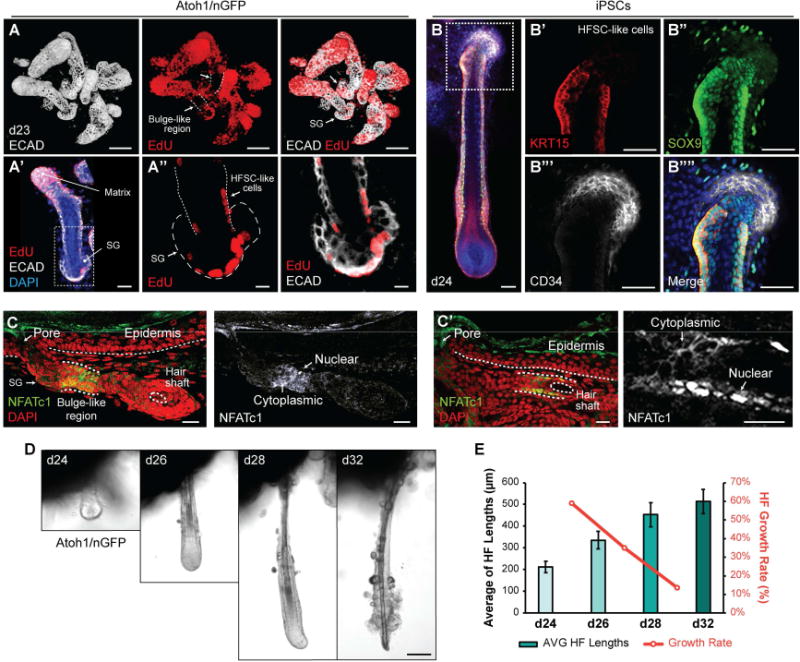
A–A″, Representative images of HFs on day 23, which were incubated with EdU solution for 24 hours. EdU incorporation was noted in the HF matrix (A), in the outer root sheath, and the SG (A′, A″). Arrows indicate HF bulge-like region. SG; sebaceous gland, HFSC; hair follicle stem cell. B–B″″, KRT15+ SOX9+ CD34− HFSC-like cells at early postnatal stage of maturity were present in the HF bulge-like region on day 24. C, C′ Representative IHC images of NFATc1 in the bulge-like region of two different Atoh1/nGFP-derived HFs. NFATc1 expression localized to the cytosol or nuclei of cells within the bulge-like region. D, E, HF morphologies were tracked during days 24–32 (D) after embedding aggregates in Matrigel on day 20 to fix aggregate position. Nine individual HFs were tracked to measure HF lengths, and HF growth rates were analyzed (E). HFs grew until day 32 while the growth rates of each interval were decreasing (D, E). HFs appeared to undergo degeneration starting on day 28 with protruding dermal papilla morphology, and eventually loosing integrity of the cells at the hair bulb region by day 32 (D). Dash-lined boxes (A′, B) indicate the area of magnification. Bars denote ± SEM. Scale bars, 100 µm (A, D), 50 µm (B–B″″), 30 m (A′, C, C′), 10 µm (A″).