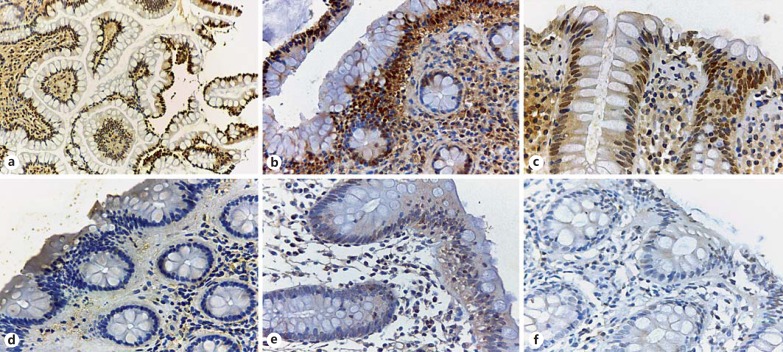
Fig. 1.
FXR immunohistochemical staining of patients with microscopic colitis and controls. a Normal terminal ileum from a control patient: there is strong nuclear staining in the epithelial cells in the villi (×40). b Right colon biopsy from a control patient; a strong nuclear positivity at the surface and upper part of the colonic glands with a gradual loss of expression in the crypts is seen (×200). c Left colon biopsy of a control patient displaying strong FXR nuclear expression (×400). d Right colon of a patient with collagenous colitis: absent FXR nuclear expression is observed (×200). Right colon (e) and left colon (f) in a patient with lymphocytic colitis displaying only minimal or no FXR nuclear staining (×400).

