FIG 5 .
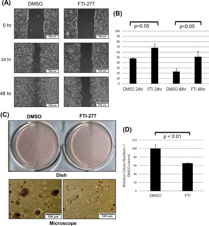
Inhibition of cellular farnesylation with FTI-277 reduces migration and colony formation of EBV-positive epithelial cells. 293EBV cells were treated with DMSO or farnesyltransferase inhibitor FTI-277 (5 μM). (A and B) Results of an in vitro wound-healing assay show that treatment with FTI-277 inhibits motility of EBV-positive epithelial 293 cells. Confluent monolayers of 293EBV cells were scraped with a plastic pipette tip, and migration of cells was analyzed. (A) Typical wounds examined under a microscope at 0, 24, and 48 h are shown (bars; 500 μm). (B) The widths of the “wounds” (scratched areas) at 24 and 48 h were measured by the use of ImageJ software. The percentage of the wound area was calculated by the following formula: wounded area (%) = (width after 24 h or 48 h/width at beginning) × 100%. The graph shows percentages of wound areas at 24 h and 48 h normalized to 0 h as 100% (means ± SD; n = 3 independent experiments [Student’s t test]). (C and D) The colony-forming assay was performed by seeding equal amounts of cells of different sets in soft agar in 6-well plates in triplicate. Numbers of colonies with a diameter greater than 200 μm were quantified after 10 days. (C) (Top panel) Dishes of colonies in soft agar. (Bottom panel) Colonies under the microscope. Bars, 500 μm. (D) Numbers of colonies per field were calculated. The graphs show relative colony numbers in control and FTI-277-treated dishes (means ± SD; n = 3 independent experiments [t test]).

