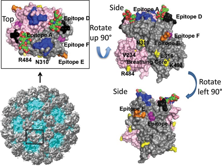
FIG 5 .

Residues important for mediating GII.4 norovirus antigenicity. A homology model of a P2 domain dimer (light gray and magenta monomers) of GII.4.2006a bound to A antigen (red and green) with identified blockade antibody epitopes A (blue), D (black), E (orange), and F (purple) and the “breathing core” residues (NERK plus residue 234) that mediate global particle conformation (yellow) color coded. The full GII.10 VLP, in which P dimers are shown in gray, is shown for context.
