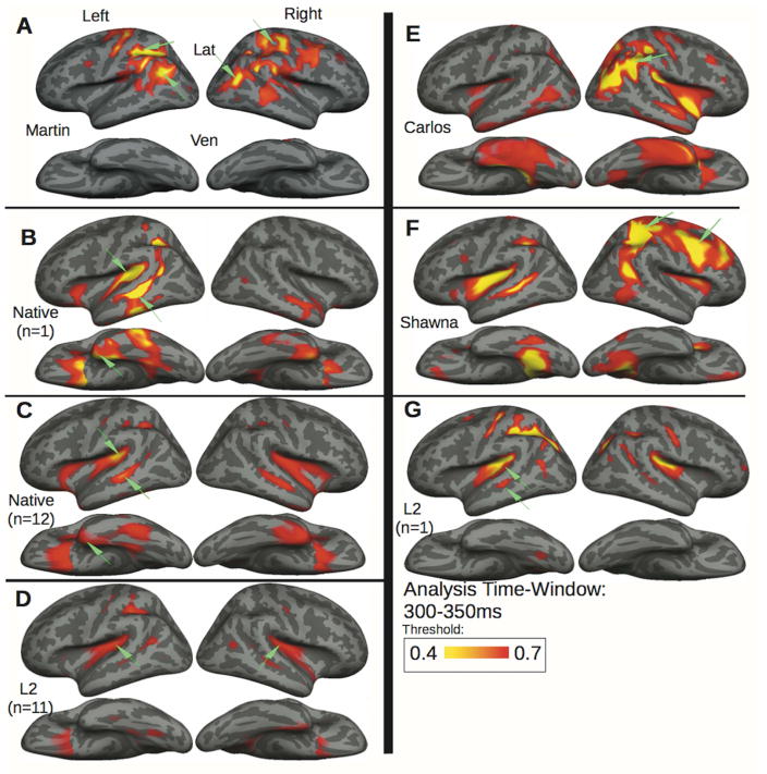
Figure 2. Semantic activation patterns to picture-primed signs.
exhibited by Martin compared with the adolescent learners Carlos, Shawna, and a representative deaf native signer and hearing L2 signer, along with the deaf native signer and hearing L2 control group averages (see Table 1). During semantic processing (300–350ms), Martin (A) shows the strongest effect in the occipito-parietal cortex bilaterally similar to those shown in the right hemisphere by Carlos (E) and Shawna (F), who shows additional left superior temporal and right frontal activity. A representative deaf native signer (B) and hearing L2 signer (G) both exhibit semantic effects in left fronto-temporal language similar to the control group averages for the native signers (C) and the L2 signers (D).

