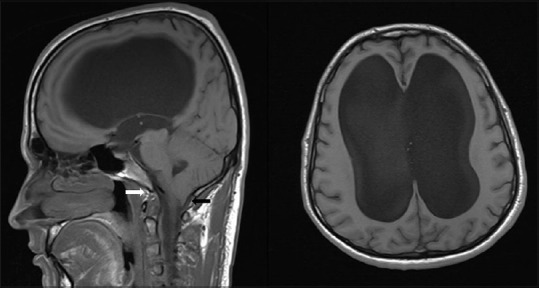
Figure 1.

T1-weighted magnetic resonance imaging: Width of lateral ventricle and the third ventricle were found to be severely wide (hydrocephalus). Hernia of cerebellar tonsils was found to be 8 mm long (black arrow), detected from foramen magnum (white arrow) to inferior (Arnold–Chiari malformation 1)
