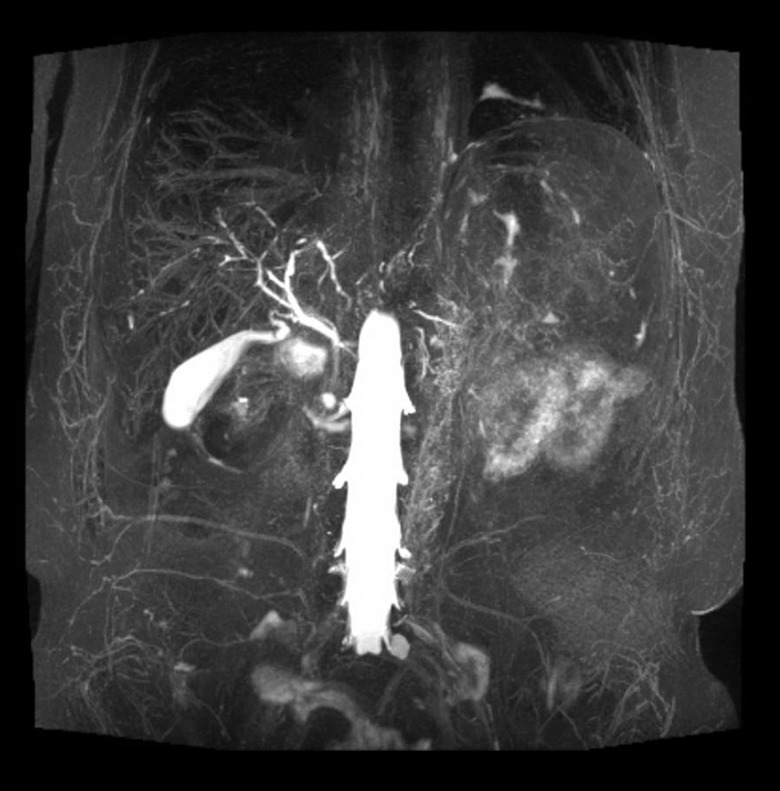
Figure 2.
The aortic wall is diffusely thickened. There is marked edema as noted on T2 STIR sequences as well as increase in signal on high B-value consistent with edema. On post-contrast administration, there is interval increase in the enhancement of the aortic wall suggestive of active vasculitis.

