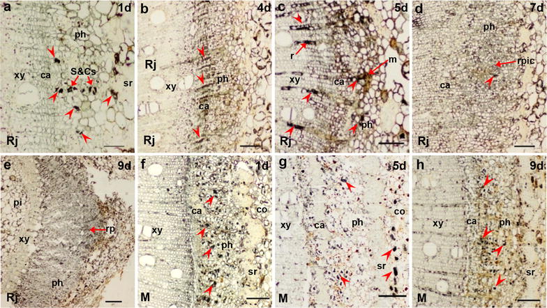
Fig. 6.

ZR immunolocalization in the stem bases of rejuvenated and mature soft shoots. a–e Transverse sections of rejuvenated soft shoots in walnut. a On 1 day of induction, ZR signals were located mainly in S&Cs. b By 4 days, as the cambium thickened, ZR became mainly concentrated in the phloem and cambium, particularly along ray cells. c By 5 days, some meristem cells had divided and the ZR signals were distributed among meristems, S&Cs and ray cells. d By 7 days, ZR signals were more evident in the initial cells of the root primordia, but less evident in cambium and S&Cs. e ZR signals were widely distributed in the phloem and root primordia by 9 days. f–h ZR was present more evident in S&Cs, but not so much in cambium of mature soft shoots on day 1 (f) and 5 (g). f ZR signals were observed between the xylem and the cambium by 5 days. g ZR signals were observed in the cortex by 5 days. h ZR signals were observed in ray cells by 9 days. The arrows indicate the ZR signals. Arrowheads indicate the tissue structure. ca cambium, co cortex, m meristem, ph phloem, pi pith, r ray cell, rp root primordia, rpic root primordia initial cells, S&Cs sieve and companion cells, sr sclerenchyma, xy xylem. Scale bars: 200 μm (e); 100 μm (b–d, f–h); and 50 μm (a)
