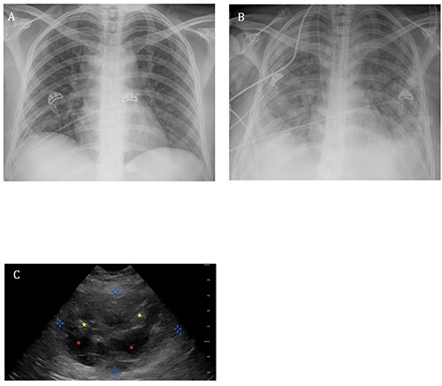
Figure 1.

A) The patient’s portable chest radiograph on admission showing subtle prominence of the interstitial markings and multiple ill-defined nodular opacities. B) Portable chest radiograph taken following endotracheal intubation demonstrating interval development of diffuse bilateral interstitial and alveolar opacities as well as diffuse granular opacity and possible right pleural effusion. C) Ultrasonographic image of the liver obtained at the bedside showing a large hepatic mass-like lesion demarcated by the blue calipers with both cystic (red stars) and solid (yellow stars) components.
