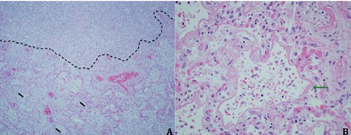
Figure 2.

A) Section of lung tissue obtained at autopsy demonstrating destruction of lung parenchyma with an abscess (above dotted black line) adjacent to an area with preserved alveolar structures and intra-alveolar neutrophils (black arrows) (hematoxylin & eosin, original magnification x 200). B, Close-up of alveoli lined with fibrinous, eosinophilic material (green arrow) consistent with hyaline membranes (hematoxylin & eosin, original magnification x 400).
