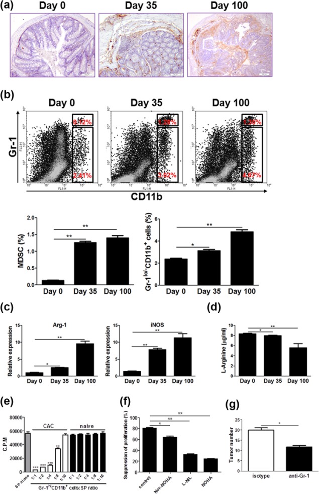
Figure 2.
MDSCs accumulate increasingly in CAC lesions and have a critical role in CAC development. On days 0, 35, and 100 after initiation of CAC induction, the following experiments were performed. (a) Colon tissues were dissected and fixed with formalin, then processed conventionally. MDSCs were detected by staining with anti-Gr-1 antibody (brown). The representative images from three independent experiments were shown. Scale bar: 200 µm. (b) Immune cells in the LP of the colon were isolated and the percentages of MPs (Gr-1lo/−CD11b+) and MDSCs (Gr-1hiCD11b+) were examined by flow cytometry. The representative plots from three independent experiments were shown. (c) LP immune cells were isolated as described in “Materials and methods.” Gr-1+CD11b+ cells in LP were purified by FACS sorting. Arginase 1 (Arg-1) and inducible nitric oxide synthase (iNOS) expression in Gr-1+CD11b+ cells was determined by quantitative RT-PCR. (d) The pieces of colon tissues were dissected and cultured in serum-free medium ex vivo for 24 h. The supernatants were collected and l-arginine contents were examined by HPLC analysis. (e) Gr-1+CD11b+ cells were isolated from the colonic LP of CAC-bearing mice and naïve littermates and cocultured with splenocytes (SP) from naïve C57BL/6 mice at titrated ratios indicated, with stimulation of Con A (5 µg/mL) for 72 h. The proliferation was evaluated by [3H]-thymidine incorporation. The data were pooled from three independent experiments. (f) The coculture of Gr-1+CD11b+ cells was isolated from the colonic LP of CAC-bearing mice and splenocytes at 1:4 ratios were preincubated with Nor-NOHA (50 µM), L-NIL (5 µg/mL) and NOHA (100 µM), respectively, and stimulated with Con A (5 µg/mL) for 72 h. The proliferation was evaluated by 3[H]-thymidine incorporation. (g) Mice subjected to CAC induction were injected intraperitoneally with anti-Gr-1 antibody or isotypes according to the regimen as described in “Materials and methods.” Tumor number in the colon and rectum was counted. The data were pooled from three independent experiments. Each group consists of six to eight mice.
*P < 0.05; **P < 0.01; ***P < 0.001 versus controls.

