Figure 3.
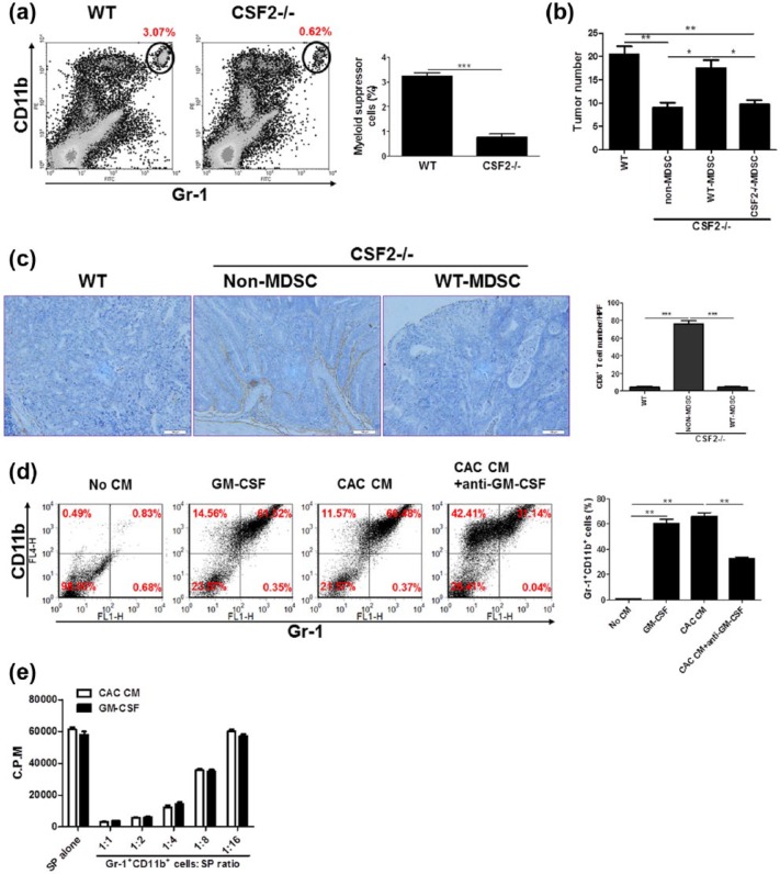
The CAC-promoting role of GM-CSF is via inducing/recruiting MDSCs. (a) CAC was induced in CSF2−/− mice and wild-type littermates and the percentages of Gr-1+CD11b+ cells in the LP of the colon were examined by flow cytometry. The representative plots from three independent experiments were shown. (b and c) Wild-type and CSF2−/− mice were transfused intravenously with MDSCs or non-MDSC (3 × 10⁶/mouse), respectively, followed by CAC induction. Tumor number in the colon and rectum was counted. (b) Colon tissues were dissected and processed conventionally. CTLs were detected by staining with anti-CD8α antibody (brown). The representative images from three independent experiments were shown. (c) Scale bar: 100 µm. (d) c-kit+lineage- hematopoietic progenitors isolated from bone marrow of C57BL/6 mice were treated with the supernatants of colon tissues of CAC-bearing mice (CAC CM) and recombinant murine GM-CSF (50 ng/mL) for 7 days. Neutralizing anti-GM-CSF monoclonal antibody (1 µg/mL) was added into the culture. The percentages of Gr-1+CD11b+ cells were examined by flow cytometry. The representative plots from three independent experiments were shown. (e) Gr-1+CD11b+ cells were pooled from CAC CM or GM-CSF-treated culture outputs and cocultured with splenocytes (SP) from naïve C57BL/6 mice at titrated ratios indicated, with stimulation of Con A (5 µg/mL) for 72 h. The proliferation was evaluated by [3H]-thymidine incorporation. The data were pooled from three independent experiments. Each group consists of six to eight mice.
*P < 0.05; **P < 0.01; ***P < 0.001.

