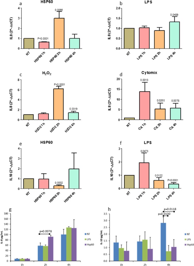
Figure 1.
IL-8 and IL-10 mRNA and protein expression after single challenges. IL-8 mRNA in 16-HBE cells treated with (a) eHSP60, (b) lipopolysaccharide from P. aeruginosa (LPS), (c) H2O2, or (d) cytomix (IL-1β+TNFα+IFNγ) (CX). (e) Treatment with eHSP60 down-regulated IL-10 mRNA at 2 h. (f) The treatment with LPS, up-regulated transitorily IL-10 expression at 1 h after exposure, which was followed by down-regulation at 2 and 4 h after exposure. (g) eHSP60 stimulation of 16-HBE cells up-regulated IL-8 protein secretion after 2 h of exposure. (h) IL-10 protein secretion was down-regulated by both LPS and eHSP60 stimulations after 4 h of exposure. Data are presented as mean ± SD of quadruplicate experiments both for mRNA and secreted proteins. The expression of all genes studied were normalized to GAPDH levels in each sample to determine the difference in expression between treated and non-treated cells using the 2−ΔΔCt method (see the “Materials and methods” section); NT indicates not treated cells.

