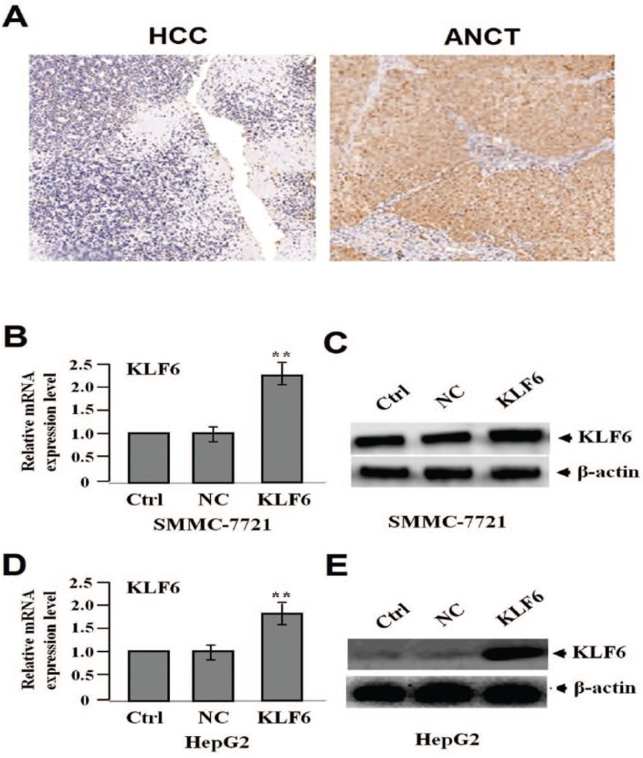
Figure 1.
The protein expression of KLF6 in HCC tissues (200). (a) HCC tissues were immunohistochemically stained with the anti-KLF6 antibody. The positive expression of KLF6 was decreased in HCC tissues compared with the adjacent non-cancer tissues (ANCT). After HCC cell lines (SMMC-7721 and HepG2) were infected with lentivirus-mediated KLF6 overexpression for 24 h, the expression of KLF6 was examined by real-time PCR (b, d) and western blot assays (c, e) (**P <0.01).

