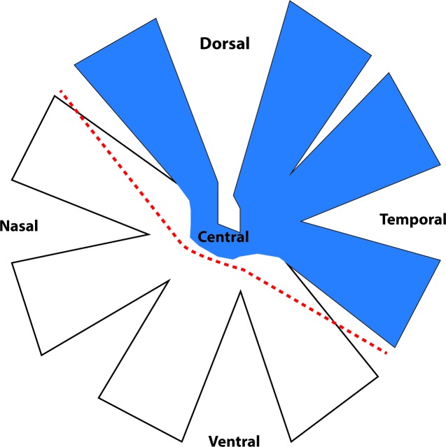
Fig 1. Schematic diagram of dissection of retina for microarray studies.
RGC somata whose axons are transected after PT of the optic nerve are located in the dorsal, temporal and central areas (indicated in blue), leaving RGCs in nasal and ventral retina initially intact but vulnerable to secondary degeneration[26]. The dashed line indicates where dorsal and ventral retinal tissue were separated.

