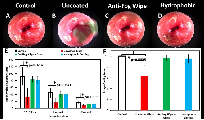
Fig 5. Representative cervical mannequin images misted with green food dye with varying treatments. with qualitative image quality assessed by 3 blinded clinicians, and quantitative image quality by computing Weber’s contrast.
The cervix mannequin has acetowhitened lesions at 12, 3, and 7 o’clock positions and a cyst at the 4 o’clock position. Control untreated glass optical window (A), dye misted untreated glass optical window (B), dye misted anti-fog wipe treated glass window (C), and dye misted hydrophobic window (D). (E) Weber’s contrast values were calculated for each lesion (n = 3) and the mean and standard deviation were determined from 3 repeated images of the same cervix. The uncoated glass (red) performed significantly worse than the control (white) at all 3 lesion positions (p<0.02 for all using 2-sample t-test and one-way ANOVA). The anti-fog wipe (green) and hydrophobic window (light blue) were not significantly different from the control group (p>0.1). (F) Qualitative assessment by 3 blinded clinicians of each treatment and control group noted a significant degradation in image quality for the uncoated glass that was misted with dye when compared to control (p<0.005).

