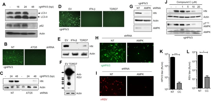
Fig 7. TDRD7 inhibits HPIV3 and RSV replication by anti-AMPK activity.
(A) HeLa cells were infected with rgHPIV3 (moi:1) and analyzed for LC3 and p62 by immunoblot. (B, C) HT1080 cells stably expressing shRNA against ATG5 were infected with rgHPIV3 (moi:1); GFP at 24 hpi (B) and viral HN protein (C) expression were analyzed at the indicated time. (D, E) HeLa cells expressing V5.TDRD7 were infected with rgHPIV3 (moi:1), GFP (D) and viral HN protein (E) expression were analyzed at 24 hpi. IFN-β pre-treatment was used as a positive control. (F) HeLa cells expressing V5.TDRD7 were infected with RSV (moi:1) and viral protein expression was analyzed at 48 hpi. Arrows indicate the polyclonal serum detecting various viral proteins. (G) HeLa cells expressing AMPK shRNA were infected with rgHPIV3 (moi:1) and viral protein (HN) expression was analyzed by immunoblot at 24 hpi. Lower panel indicates the AMPK levels in these cells. (H) HeLa cells expressing AMPK shRNA were infected with rgHPIV3 (moi:1) and GFP expression was analyzed by fluorescence microscopy at 24 hpi. (I) HeLa cells expressing AMPK shRNA were infected with rrRSV and virus-encoded red fluorescent protein expression was analyzed by fluorescence microscopy at 24 hpi. (J) HeLa cells were pre-treated with various concentrations of Compound C for 1h, and then infected with rgHPIV3. Viral protein (HN) and pAMPK (Thr172) levels were analyzed at 24 hpi by immunoblot. (K, L) HeLa cells were pre-treated with Compound C (CC, 10 μM) for 1h, and then infected with rgHPIV3 (K) or rrRSV (L). Infectious virus particle release in the culture supernatants was analyzed by fluorescence focus assay (expressed in ffu/ml). NT, non-targeting, EV, empty vector, * indicates p<0.05. The results presented here are representatives of at least three biological repeats.

