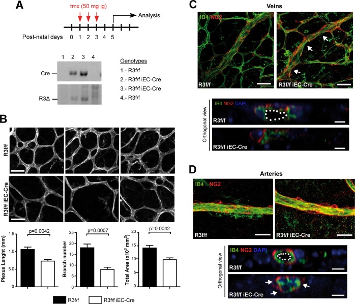
Fig 2. Endothelial deletion of Rasa3 results in defects in retinal vascularization.
A. Experimental design of deletion of Rasa3 in R3f/f and R3f/f iEC-Cre pups via tamoxifen (tmx) intragastric (ig) injections at P1, P2 and P3. At P5, the R3∆ allele was only detected in Cre-positive R3f/f newborns. Lower panels: genotyping of 4 mice by PCR for the Cre transgene (above) and the Rasa3∆ allele (below) detection. When the Cre transgene is present, the Rasa3∆ allele appears, indicating the deletion of exons 11 and 12 of the Rasa3 gene. The genotype of the 4 mice is indicated on the right. B. Immunofluorescence analysis of R3f/f and R3f/f iEC-Cre retinal plexus stained for the IB4 endothelial cell marker (upper images). Representative images of 4 independent experiments are shown. Bars = 50 μm. (Graphs) Quantification of cumulative length (left), number of branches (center) and area (right) of retinal vascular plexuses from tamoxifen-treated R3f/f and R3f/f iEC-Cre newborns. Data are represented as mean ± SEM. C. Immunofluorescence analysis of retinas from R3f/f and R3f/f iEC-Cre newborns using an endothelial (IB4, green) and a pericyte (NG2, red) marker. Representative images of twisted regions (arrows) in R3f/f iEC-Cre veins are shown. (Lower panel) Orthogonal reconstructions of confocal Z-stack in one representative R3f/f iEC-Cre vein showing luminal occlusion. Nuclei were stained with DAPI (blue). D. Representative images of arteries in retinas of R3f/f and R3f/f iEC-Cre pups. (Lower panel) Orthogonal reconstructions of confocal Z-stack in one representative R3f/f iEC-Cre artery with luminal occlusion. The lumen is outlined with a white dotted line in the control. Bars are 50 μm. The p values are shown (Unpaired t-test).

