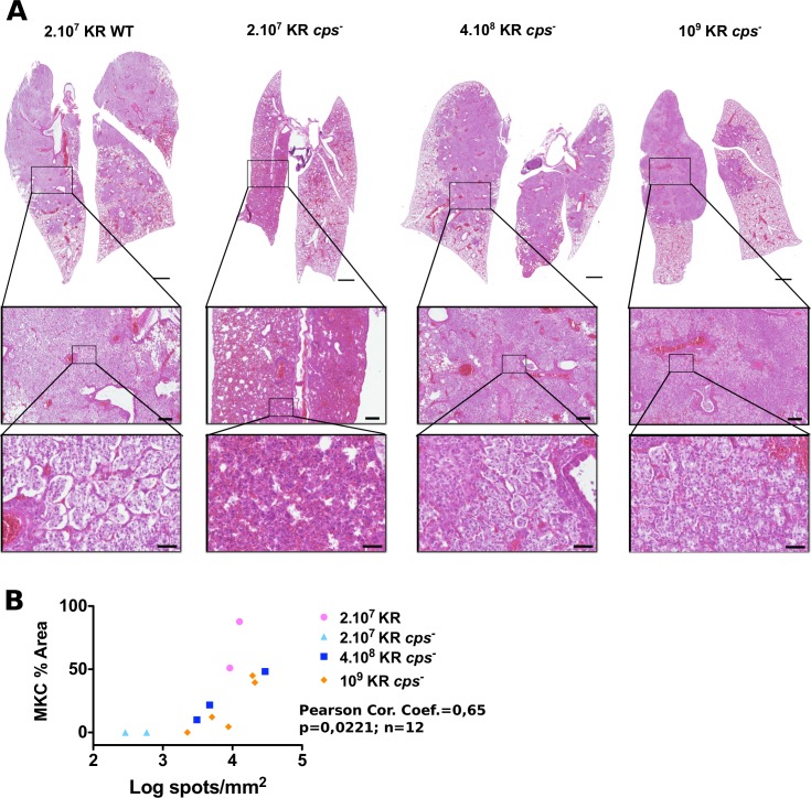
Fig 3. Histology of BALB/c lungs infected by wild-type K. rhinoscleromatis and KR cps-.
(A) Lungs of mice infected with 2.107 KR WT, 2.107 KR cps-, 4.108 KR cps-, and 109 KR cps- were resected 4 days post-infection and examined by histology. Lungs infected with 2.107 KR WT (left) presented the classical pattern characterized by many alveoli, with an intact epithelial layer, filled with Mikulicz cells. On the contrary, lungs infected with 2.107 KR cps- (middle left) showed dense inflammatory infiltrate containing numerous polymorphonuclear cells and absence of Mikulicz cells. Lungs of mice infected with 4.108 KR cps- (middle right) or 109 KR (right) showed the presence of Mikulicz cells similarly to wild-type infection. Insets show magnification of representative zones of the lungs. Scale bars are 1 mm (top), 200 μm (middle row) and 50 μm (bottom). Images are representative of 4 (KR WT), 4 (2.107 KR cps-), 4 (4.108 KR cps-) or 5 (109 KR cps-) mice from 2 to 3 independent experiments. (B) Correlation between the area covered by Mikulicz cells and number of bacteria spots in lungs sections of mice at 96 hours after infection with 2.107 KR WT, 2.107 KR cps-, 4.108 KR cps-, 109 KR cps-.

