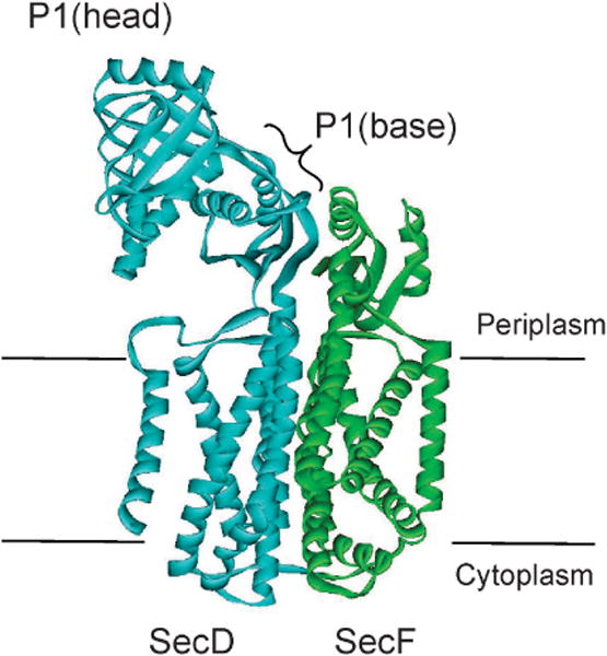
Figure 10. Structure of SecDF.

The structure of T. thermophilus SecDF, which is encoded as a single polypeptide chain, is colored to represent the individual SecD and SecF polypeptides found in E. coli. Transmembrane helices 1 – 6 (blue) represent E. coli SecD. The periplasmic P1 domain between TM1 and TM2 is shown at the top of the figure. The head and the base subdomains are indicated. Transmembrane helices 7 – 8 represent E. coli SecF.
