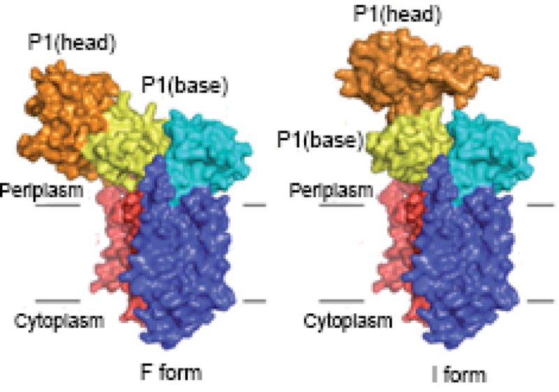
Figure 11. Two conformations of SecDF.

The P1 domain of SecDF, shown extending into the periplasm comprises two subdomains: a P1 head (orange) and P1 base (blue). The protein was crystallized in the F form (left hand side) with the P1 domain positioned so that the head is bent toward the membrane. The I form shows the head directly above the base. This structure is a model built from superimposing the base subdomain of the isolated P1 structure onto that of the full-length SecDF. (used with permission from Tsukazaki et al. (233)
