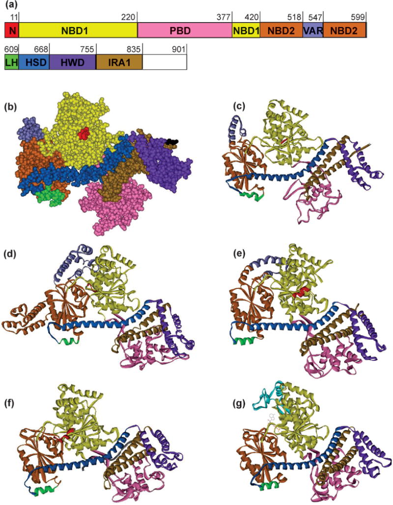
Figure 4. Structures of SecA monomers.

(a) The sequence is E. coli SecA with the domains colored as in the structures. See text for domain abbreviations. (b) SecA from E. coli in CPK representation, PDB 2FSF with the PBD modeled in based on B. subtilis SecA, PDB ITF5, by A. Economou. (c) Ribbon representation of E. coli SecA shown in (b). (d) – (g) Ribbon representation of SecA from the following species: (d) T. thermophilus, PDB 2IPC, (e) M. tuberculosis, PDB 1NL3, (f) B. subtilis, PDB 1M6N, and (g) T. maritima, PDB 3JUX.
