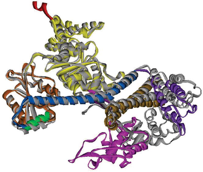
Figure 5. Comparison of the open and closed structures of SecA.

The closed conformation of B. subtilis SecA (PDB 1M6N) is shown as the gray ribbon. The open conformation of B. subtilis SecA (PDB 1TF5) is shown in ribbon representation with the domains colored as in Figure 4. The Protein Binding Domain (pink) is the only domain that has moved.
