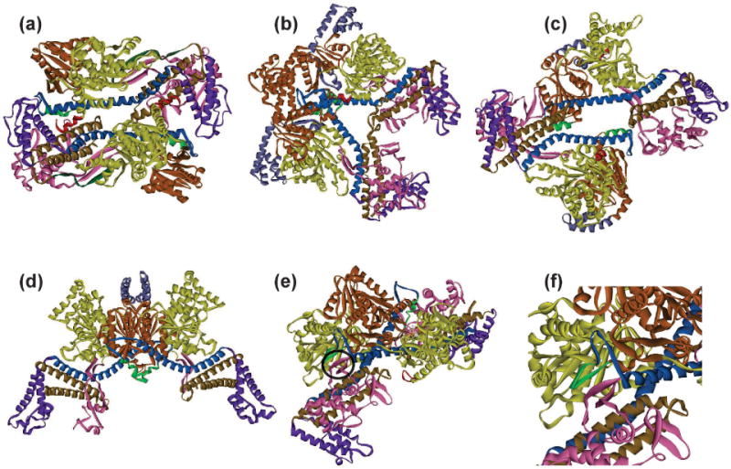
Figure 6. Dimeric forms of SecA.

Dimeric structures of SecA with the domains colored as in Figure 4. The SecA species are from: (a) B. subtilis PDB 1M6N, (b) T. thermophilus PDB 2IPC, (c) M. tuberculosis PDB 1NL3, (d) E. coli PDB 2FSF, (e) B. subtilis PDB 2IB; the three-stranded β sheet that forms the interface is circled and enlarged in (f).
