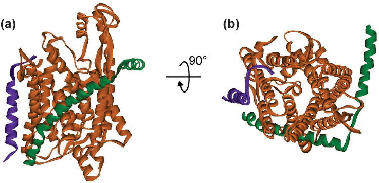
Figure 7. Structure of SecYE.

The structure of SecYEβ (PDB 1RHZ) is shown as an example of the common structure of the SecYE core. SecY is shown as the orange ribbon, SecE as the green ribbon and Secβ (SecG in E coli) as purple. The view in (a) is in the plane of the membrane with the cytoplasmic face at the top and the periplasmic face at the bottom. The view in (b) results from a 90° rotation toward the viewer to show the channel in the translocon from the cytoplasmic face. The plug can be seen in the middle of the channel at the periplasmic side.
