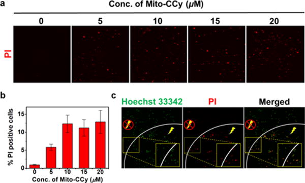
Figure 8.

Effect of NIR irradiation following Mito-CCy administration on the viability of HeLa cells. (a) Confocal fluorescence images of propidium iodide-treated HeLa cells after incubation with various concentration of Mito-CCy for 4 h, following by 730 nm irradiation (2.3 W/cm2) for 10 min. (b) The proportion of PI-positive cells was determined from the ratio of PI-positive cells to the total number of cells as determined by Hoechst 33342 staining. (c) Confocal fluorescence images of HeLa cells obtained after incubation with Mito-CCy (20 μM) for 4 h following 730 nm laser irradiation (2.3 W/cm2) for 10 min. Dead cells are labeled in red by PI staining, whereas all cells were visualized using Hoechst 33342 (green). A Ti:Sa femtosecond-pulsed laser (Chameleon XR by Coherent, 200 fs pulse width, 90 MHz repetition rate) was used. The energy density (fluence) was 1380 J/cm2.
