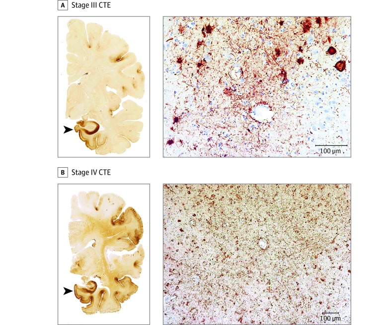
Figure 2. Representative Images of Phosphorylated Tau Pathology at CTE Pathological Stages III and IV.
CTE indicates chronic traumatic encephalopathy; NFT, neurofibrillary tangle; ptau, phosphorylated tau. For all images, 10-µm paraffin-embedded tissue sections were immunostained with microscopic mouse monoclonal antibody for phosphorylated tau (AT8) (Pierce Endogen). Positive ptau immunostaining appears dark red, hematoxylin counterstain; calibration bar indicates 100 µm. In stage III CTE, multiple CTE lesions and diffuse neurofibrillary degeneration of the medial temporal lobe are found. In stage IV CTE, CTE lesions and NFTs are widely distributed throughout the cerebral cortex, diencephalon, and brain stem. All hemispheric tissue section images are 50-µm sections immunostained with mouse monoclonal antibody CP-13, directed against phosphoserine 202 of tau (courtesy of Peter Davies, PhD, Feinstein Institute for Medical Research; 1:200); this is considered to be an early site of tau phosphorylation in NFT formation. Positive ptau immunostaining appears dark brown. A, Former NFL player with stage III CTE. There are multiple large CTE lesions in the frontal cortex and insula; there is diffuse neurofibrillary degeneration of hippocampus and entorhinal cortex (black arrowhead). Perivascular CTE lesion: a dense collection of NFTs and large dot-like and threadlike neurites enclose several small blood vessels. B, Former NFL player with stage IV CTE. There are large, confluent CTE lesions in the frontal, temporal, and insular cortices and there is diffuse neurofibrillary degeneration of the amygdala and entorhinal cortex (black arrowhead). Perivascular CTE lesion: a large accumulation of NFTs, many of them ghost tangles, encompass several small blood vessels.

