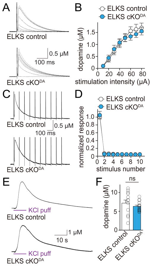
Figure 4. ELKS1α and ELKS2α are dispensable for dopamine release in the dorsal striatum.
(A) Sample traces of dopamine release evoked by electrical stimulation in slices of ELKS cKODA and sibling ELKS control mice. In all electrophysiological experiments, ELKS cKODA are mice in which ELKS1α and ELKS2α are removed specifically in dopamine neurons, and ELKS control mice are siblings of ELKS cKODA that have one wild-type allele for each ELKS encoding gene.
(B) Quantification of peak amplitude in (A). ELKS control n = 12 slices/4 mice, ELKS cKODA 12/4 (p = 0.53 for genotype, p < 0.001 for stimulation intensity and p = 0.92 for interaction; two-way ANOVA).
(C) Sample traces (average of 4 sweeps) of dopamine release evoked during 10 Hz electrical stimulation in ELKS control and ELKS cKODA mice.
(D) Quantification of (C). Amplitudes were normalized to the average first amplitude in ELKS control. ELKS control n = 12 slices/4 mice, ELKS cKODA n = 12/4 (p = 0.96 for genotype, p < 0.001 for stimulus number and p = 0.96 for interaction; two-way ANOVA).
(E) Sample traces of dopamine release evoked by a local puff of 100 mM KCl for 10 s in ELKS control and ELKS cKODA mice.
(F) Quantification of peak amplitude in (E). ELKS control n = 13 slices/5 mice, ELKS cKODA n = 15/5.
All data are mean ± SEM. ns, not significant; two-way ANOVA for (B, D) and Mann-Whitney rank sum test for (F).

