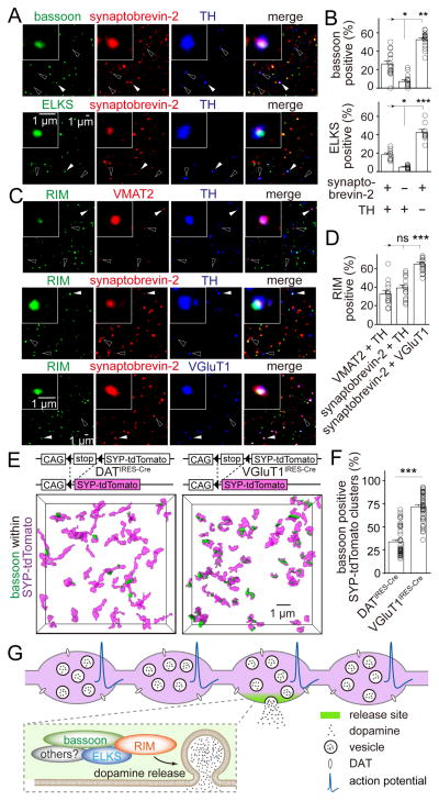
Figure 7. Many dopamine varicosities lack active zone-like release sites.
(A) Representative confocal images of sparsely plated striatal synaptosomes stained for synaptobrevin-2, TH and bassoon (top) or ELKS (bottom). Filled arrowheads indicate synaptosomes containing all three markers. Hollow arrowheads indicate synaptosomes containing synaptobrevin-2 and TH but not bassoon or ELKS.
(B) Quantification of (A) showing the percentage of particles that contain bassoon or ELKS. Each circle represents the average result of an area with 1,500–6,000 synaptosomes. n = 18 areas/3 mice for bassoon and 9/3 for ELKS.
(C) Striatal synaptosomes stained for RIM, VMAT2 and TH (n = 14/3); RIM, synaptobrevin-2 and TH (n = 17/3); or RIM, synaptobrevin-2 and VGluT1 (n = 17/3). Filled arrowheads indicate synaptosomes (insets) containing both markers (VMAT2 and TH, synaptobrevin-2 and TH, or synaptobrevin-2 and VGluT1) and RIM. Hollow arrowheads indicate synaptosomes containing the same markers but not RIM.
(D) Quantification of (C) showing the percentage of particles that contain RIM. n = 14 areas/3 mice for VMAT2 and TH positive synaptosomes, 17/3 for synaptobrevin-2 and TH positive synaptosomes and 17/3 for synaptobrevin-2 and VGluT1 positive synaptosomes.
(E) Representative surface rendered 3D-SIM images of bassoon within vesicle clusters in the striatum. Vesicles in dopaminergic neurons or in glutamatergic neurons were labeled by crossing Cre-dependent synaptophysin-tdTomato mice (SYP-tdTomato) with DATIRES-Cre or VGluT1IRES-Cre mice, respectively.
(F) Quantification of (E) showing the percentage of vesicle clusters associated with bassoon. SYP-tdTomato x DATIRES-Cre n = 49 regions/4 mice, SYP-tdTomato x VGluT1IRES-Cre n = 46/3.
(G) Current model of cellular and molecular architecture for dopamine secretion. Most dopamine varicosities contain clusters of dopamine vesicles and DAT along dopamine axons, but only ~30% of them contain active zone-like release sites composed of the scaffolds bassoon, RIM, ELKS and likely other active zone proteins including Munc13. When action potentials propagate through dopamine axons, dopamine vesicles fuse at these sites with a very high release probability, and fusion requires the presence of RIM. This architecture generates a fast, local rise in dopamine followed by a rapid decay, which may support fast dopamine coding.
All data are mean ± SEM. *** p < 0.001, ** p < 0.01, * p < 0.05, ns, not significant; Kruskal-Wallis analysis of variance with post hoc Dunn’s test for (B, D), Mann-Whitney rank sum test for (F).

