
Figure 7.
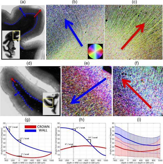
Fibers typically curve more at the sulcal wall than they do at gyral crowns. A histological slice containing the SFG (a) is shown, along with the WMGM border (blue) and the normal to the border (yellow lines). Arrows are shown at the wall (blue) and crown (red) going from white matter into gray matter. Both arrows are perpendicular to the WMGM boundary. High‐resolution HSB images in the same places at the wall b) and crown (c) demonstrate the high curvature of fibers entering the cortex at the walls (b) and the long, straight fibers at the crown (c). A myelin‐stained slice containing IFG and FOG regions (d), and high‐resolution HSB images at the wall (e) and crown (f), show similar trends. The fibers from (a) to (c) are tracked from white matter, into gray matter, and the angle these fibers make with the normal to the WMGM border is recorded (g) for the wall (blue) and crown (red). The slope of this curvature is marked in various locations. The fibers from (d) to (f) are similarly tracked, and the angle at these locations from the wall (blue) and crown (red) are plotted (h). Finally, the results from all gyral blades analyzed (i) are shown for the crown (red) and wall (blue) with the mean (solid line) and standard deviations (vertical lines) plotted for each [Color figure can be viewed at http://wileyonlinelibrary.com]